If work in the field of T-cell immunology, these scenarios probably do not appear foreign to you these days.
Scenario 1: You ran scRNA-Seq/CITE-Seq combined with scVDJ-Seq on fresh tumor sample. From the UMAP, you identified clusters of T cells that appear to be activated or exhausted, with strong signal of clonal expansion. On the other hand, you know the dozens of neoantigens and tumor-associated antigens that these T cells may react to. Understanding whether these T cells indeed react to the tumor cells would greatly enhance our understanding of disease progression and treatment response.
Scenario 2: You dosed the patients with cancer vaccines. From PBMC ELISPOT you can see that the patient mounted immune responses against the antigens. Better yet, some of the antigens are shared antigens. If you can confirm the TCR:pMHC interaction, you not only confirm the mechanism of action of your vaccine on this patient, you may also have a TCR that may help other patients as TCR-T or (with additional engineering) TCR-based T cell engagers (TCR-TCEs).
Scenario 3: With the help of AI, you designed a set TCRs or TCR-mimicking binders against a highly valuable pMHC target. This sequence may be an improvement of a parental sequence you or someone else discovered using other tools, or a completely novel sequence generated by AI. Knowing the potency of this set of TCRs or TCR-mimicking binders would offer valuable stepping stones for their further improvements.
In each of these scenarios, you have the full sequences of the TCR candidates. Although you may not know exactly which MHC presents which antigen at what epitope, you at least know the patient’s HLA types and the sequence contexts of the antigens. In other words, you have perfectly testable hypotheses, but, experimentally testing them somehow seems beyond reach, or at least very inconvenient.
Why Is It Hard and Slow to Test TCR:pMHC Experimentally?
The problems and solutions with MHC
Let’s unpack the situation by first looking at the MHC of “TCR:pMHC”.
If the MHC is Class I, chances are you have looked through the the catalogs of MBL, BioLegend, Immudex, Kactus, etc. for soluble MHC multimers. But unfortunately, unless the MHC of interest is A*02:01, A*11:01, or a few other very common ones, it’s unlikely that you can find what you need. If the MHC is Class II, there’s even less chance that a validated multimer reagent is available.
How about we go beyond multimers and directly use antigen-presenting cell (APC)? Compared to simply binding to multimers, cell-based assays offer more functional and biologically relevant data anyway. You are absolutely right! But now the question is, where do you get the APC? If your lab is a molecular- or computation-focused lab, with some distance from the clinic, patient-derived B-LCLs and MDDCs are probably not available to you without significant logistical and R&D effort. Even when MDDCs are available, their quantity may be limited.
How about we express recombinant MHC in an MHC-null cell line such as K562? Yes! We are getting somewhere. This is exactly the approach RootPath takes. In fact, RootPath has a meticulous system to ensure and confirm that that all of the functional MHCs of interest are properly expressed. K562 is not the only cell line we can use. If K562 happens to express the antigen of interest (making the ’no antigen control’ hard to execute), we can use other cell lines such as HEK293, HeLa and CHO.
The problems and solutions with peptide
For now let’s temporarily consider the MHC problem solved and examine the next input: the p in “TCR:pMHC” – peptide, epitope, antigen, different granularity, same element.
In APC-based assays, the conventional method is to pulse the epitope peptide onto the APC. But we need to be careful here. We need to ensure that either the peptide is already the minimal epitope, which can be loaded to the MHC cleft without proteolytic processing, or can be properly processed by the APC. We know professional APCs like B-LCLs and MDDCs can do this. But how about K562? Well, we have a good news for you: if you expressed the antigen intracellularly with proper ER- and endosome/lysosome-targeting signal peptides, the antigen can be processed and presented efficiently, as long as the epitope binds the MHC molecules. We have used dozens of published TCR:pMHC pairs to confirm this, in both Class I - CD8 and Class II - CD4 context. In practice, we need to optimize antigen density and have other tricks up our sleeve (such as Ii/CD79-based presentation for Class II antigens). But on the high level, we can check ‘peptide’ off for now.
The problems and solutions with TCR
Finally, it’s time to welcome our main character, the TCR of “TCR:pMHC”. The gold-standard method to study the function of TCR is to make recombinant TCR-T cells, which are co-cultured with the antigen-loaded APCs and observe T cell activation or APC death at different effector-to-target (E:T) ratios. To make TCR-T cells, the conventional approach involves producing retroviral or lentiviral particles, which are then used to transduce T cells (either cell line such as Jurkat or donor T cells). If the transduction efficiency is low, CD3-based enrichment is often necessary.
It is hard to imagine how a single lab generates 100s of viral stocks and maintain 100s of TCR-T cell lines for a project featuring fast iterative cycles. Again, we have a good news for you. We discovered that TCR genes can be introduced to T cells via mRNA electroporation to achieve sufficient expression level. This not only cuts the viral production and cell line enrichment out of the loop to greatly accelerate the process, but also make the process scalable, since mRNA production, purification, quantification and electroporation can all be done in 96- or even 384-well plates. No one needs to take care of hundreds of 10 cm dishes of packing cells anymore! We have accumulated a rich body of data to prove the feasibility of this approach.
Overall Workflow
Now we can put the pieces together. Here’s the workflow of a typical TCR Decoder™ project.
Step 1: TCR Synthesis. After we receive the TCR sequences, we will construct the TCR gene as plasmids, verify their sequences and process the plasmid into a linear format ready for in vitro transcription (IVT). In parallel, we prepare MHC and antigen constructs as IVT templates as well.
Step 2: In vitro Transcription. We use an optimized, proprietary chemistry to produce modified, capped and A-tailed mRNA constructs.
Step 3: Prepare T cells. We prefer using donor T cells (as opposed to Jurkat) since their signaling pathway better mimics T cells in the natural context. We have a continuous T cell procurement, QC and onboarding process to ensure supply of qualified donor T cells. To ensure there is no interference from endogenous TCR genes, we knockout their endogenous TRA and TRB genes. The resultant T cells are then confirmed to be CD3-negative, then cryopreserved. Once a TCR Decoder™ project starts, as we prepare the genes and mRNAs, we simultaneously thaw and revive aliquots of the T cells to use.
Step 4: mRNA electroporation. This is the crown jewel of the entire process. TCR-encoding mRNAs are introduced to T cells in 96-well format to form transient-expression TCR-T cells. More importantly, as quick as 24 hours after electroporation, surface expression level of TCRs are already sufficient to test TCR function. In parallel, mRNAs encoding MHCs and antigens are electroporated into the synthetic APC (such as K562), in a scheme to ensure MHC expression and optimize antigen density (see below).
Step 5: Co-culture of TCR-T cells and antigen-loaded APC. The TCR-T cells and synthetic APC are then co-cultured in IFN$\gamma$ ELISPOT plates. After 12 hrs of co-culture, the cells are discarded and the plates are stained with IFN$\gamma$ to quantify for its level.
Example Data
Here are some example data generated by this workflow. We highlight this example because all of the candidate MHCs are Class II, and all of the candidate TCRs are from CD4 T cells. To our knowledge, our TCR Decoder™ service is the only one suitable for scalable CD4 / Class II TCR:pMHC discovery.
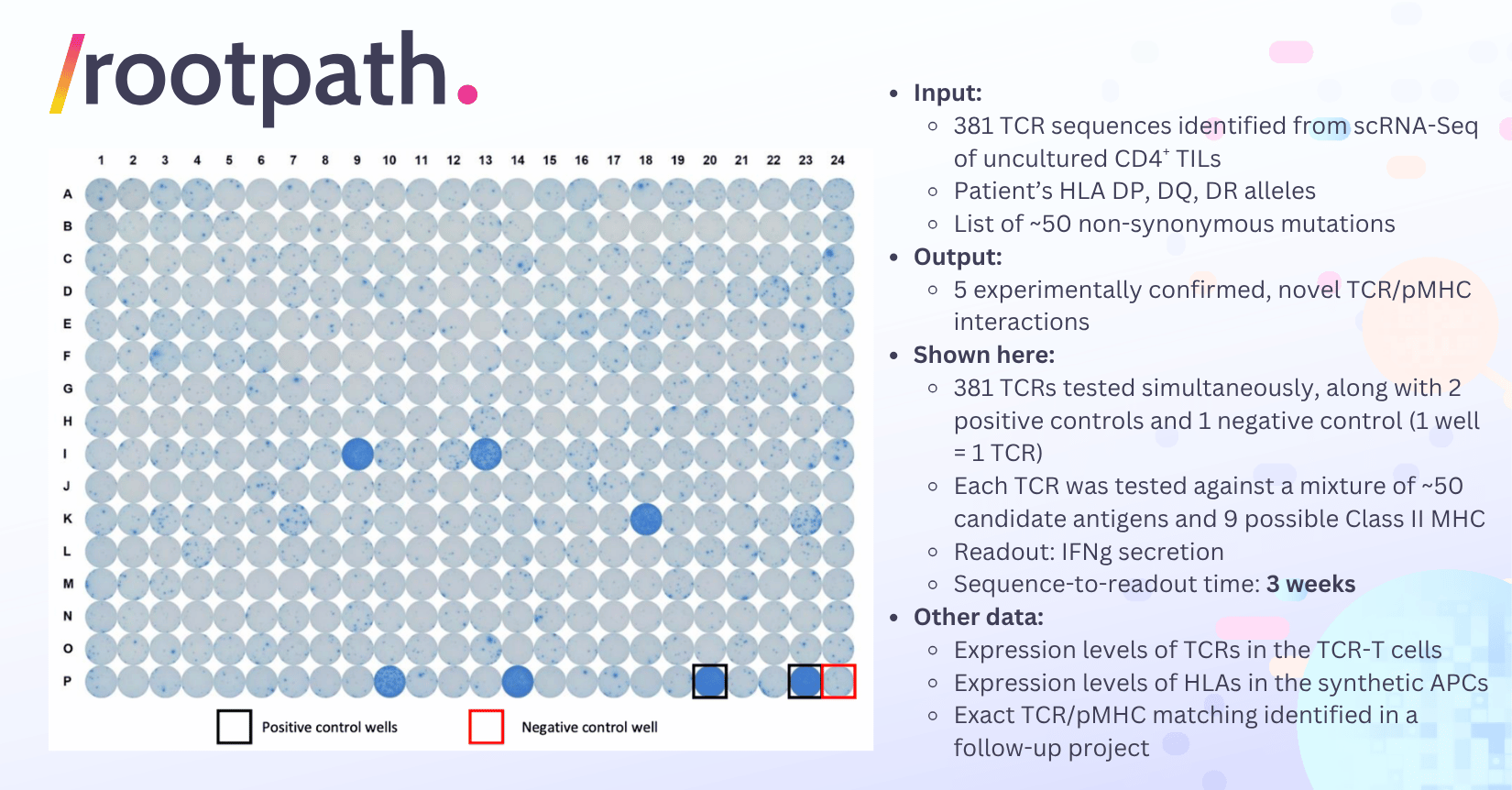
In this project, our client identified 381 $\alpha$-$\beta$ paired TCR sequences from CD4 T cells in a tumor sample via scRNA-Seq and would like to know which (if any) of them reacts to which of the Class II MHC-restricted neoantigens caused by the ~50 non-synonymous mutations within the tumor. The client sent us the TCR sequences, the HLA genotypes and the mutation information electronically.
Upon receiving the information, we synthesized the 381 TCR genes and produced 381 preps of transient-expression TCR-T cells in 96-well format using mRNA electroporation (nowadays the total time needed for gene synthesis and TCR-T production is as short as 2.5 weeks). We first measured expression level of TCR/CD3 complex of for each TCR-T line. Most showed sufficient level.
In parallel, we produced 4 MHC mRNA Groups, each group containing genes for 1 HLA-DP, 1 HLA-DQ, and 1 HLA-DR heterodimers (covering all of the 9 possible Class II HLA heterodimers for this patient, note that the patient is homozygous for some HLA alleles). We also produced one Antigen mRNA Group, containing all of the ~50 non-synonymous mutations in the format of tandem minigenes (TMGs, 10 epitopes per TMG) as well as two internal-control antigens. We prepared 4 Synthetic APC Preps, each electroporated with its corresponding MHC mRNA Group and the common Antigen mRNA Group. We confirmed the expression level of HLA-DP, -DQ and -DR on each Synthetic APC Prep, and used the internal-control pMHC to ensure that processing and presentation machinery works properly. See the section below for the concept of MHC mRNA Group and Antigen mRNA Group, and see this post for the detailed logic of HLA coverage and internal-control pMHC.
Next, we pooled all of the Synthetic APC Preps to form the “Master Synthetic APC Mixture” and added an aliquot of it to each well of a 384-well plate. Finally, we added one TCR-T prep to each well of the 384-well plate, forming 381 co-culture experiments (i.e., 1 well = 1 TCR). We included 2 positive-control TCRs each corresponding to an internal-control antigen and 1 negative control co-cultures to round up the 384 well plate.
After overnight co-culture, we detected IFN$\gamma$ secretion from each well using ELISPOT. As shown on Figure 2, both positive-control co-cultures showed strong signal, while the negative control co-culture generated low background. More importantly, 5 out of the 381 trial co-cultures showed unambiguous, positive signal. In follow-up experiments we identified the restricting elements, the exact neoantigens and confirmed the mutation-specific nature of the 5 TCR:pMHC interactions.
The Beauty of Being Digital
A key feature of RootPath’s TCR Decoder™ service is that the interface between the user and RootPath’s lab is purely digital. You only need to send the TCR sequences, patient’s HLA type and candidate antigen sequences, and get back functional data and full analysis report. The entire process doesn’t involve any shipping or custom clearance, regardless where you are in the world. A key component to ensure the digital nature is the synthetic APC expressing all patient’s MHC, which is functionally equivalent to patient-derived B-LCL and MDDC.

As shown in Figure 3, the synthetic APC is created by co-electroporation of a mixture of mRNA encoding a group of MHC molecules (referred to as MHC mRNA Group) and a mixture of mRNA encoding a group of antigens (referred to as Antigen mRNA Group). Each antigen encoding mRNA contains an N-terminal ER-targeting signal peptide, candidate antigen, a positive-control epitope, and a C-terminal endosome/lysosome-targeting signal peptide. The length of candidate antigen is up to 1 kb, which can be a long ORF (in case of viral antigen or tumor associated antigen), or 10 to 12 tandem minigenes (TMGs). Again, you can refer to this post for the detailed logic of HLA coverage and internal-control pMHC.
We have used dozens of published TCR:pMHC pairs to validate this system, and would be happy to share the data.
Closing Remarks
There are a lot of question you may have and I couldn’t cover in this post. How well do the TCRs express in donor T cells? How do we ensure the MHCs are expressed? How do we know the antigens are expressed, processed and presented? What are the positive control and negative control of each steps? What can we do to further delineate MHC and antigen after initial screening? So on and so forth.
I plan to answer these questions in future posts and update this post with links to them. But for now, let me just say that every step has been meticulously controlled and verified, and the entire workflow has been demonstrated in dozens of successfully executed projects, with customers from leading TCR labs. If you want to know now, you can always send me an email at xi@rootpath.com, or schedule an online meeting with me, where I can show you more data.
It’s my humble opinion that your powerful discovery engine finally has an experimental partner built for the digital age! Happy TCRing!
– Xi Chen