As discussed in our overview post, in our TCR Decoder™ services we bypass the reliance on patient-derived B-lymphoblastoid cell lines (B-LCL) and monocyte-derived dendritic cells (MDDC) by co-expressing the patient’ MHC genes and the antigen genes in cell lines like K562. On a high level, Figure 1 below shows the general idea. We select all or a subset of the patient’s MHC genes of interest to form an MHC mRNA Group. Similarly, we select all or a subset of the antigen of interest for form an Antigen mRNA Group. We then co-electroporate the MHC mRNA Group and the Antigen mRNA Group into a synthetic APC cell line such as K562 to form a Synthetic APC Prep. We make multiple Synthetic APC Preps to cover all MHCs and all antigens, and mix them to form a Master Synthetic APC Prep, which is co-cultured with TCR-T cells.

Many aspects, such as the molecular designs of the MHC genes and antigen genes, and how we ensure antigen density, deserve their own posts. In this post, I’d like to focus on another key aspect: how we verify all the Class I and Class II MHCs that we want to express in the Synthetic APC Preps cell are indeed expressed.
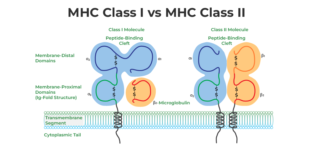
To be more specific, the overall goal in this part of the project design is to make sure that the presence of every MHC allele or heterodimer of interest can be examined on the protein level using antibodies. To better understand this goal, let’s take a step back and review the genetics of Class I and Class II MHC.
Class I MHC Genetics
Class I MHC molecule is formed by the HLA Class I $\alpha$ chain (blue in Figure. 2, left) and the ${\beta}2$-microglobulin (${\beta}2m$) molecule (orange in Figure. 2, left). Each individual has 3 Class I MHC loci in the genome: HLA-A, HLA-B and HLA-C, while each locus has 2 alleles. In this post, we refer to these alleles as HLA-A.1, HLA-A.2, HLA-B.1, HLA-B.2, HLA-C.1, HLA-C.2.
Class I MHC Surface Expression
To be able to examine every Class I MHC of interest, one sensible approach is to send mRNAs encoding in only 1 HLA-A allele, only 1 HLA-B allele, and only 1 HLA-C allele into the Synthetic APC Prep. For example:
| HLA-A | HLA-B | HLA-C | |
|---|---|---|---|
| MHC mRNA Group 1 | HLA-A.1 | HLA-B.1 | HLA-C.1 |
| MHC mRNA Group 2 | HLA-A.2 | HLA-B.2 | HLA-C.2 |
We can then use antibodies against pan-HLA-A, pan-HLA-B and pan-HLA-C to stain the MHCs. If all 3 stains are positive, we know that all of these 3 MHCs are expressed. In practice, this approach encountered 2 limitations. First, not all HLA-A alleles can be stained efficiently by the pan-HLA-A antibody (same for other isotypes). Second, as eluded to above, if we send in HLA-A.1 + HLA-B.1 + HLA-C.1, the HLA-C.1 would be outcompeted by HLA-A.1 and HLA-B.1 and not expressed.
Therefore, we revised our approach. First, we incorporated epitope tags (i.e., HA tag and Flag tag) to the N-terminus of the HLA Class I $\alpha$ chain (following the signal peptide sequence). Specifically, all alleles with suffix ‘.1’ are tagged with HA, and all alleles with suffix ‘.2’ are tagged with Flag. Second, we do not mix HLA-C with HLA-A or B. Instead, we create another Synthetic APC Prep, with HLA-C.1 (HA tagged) and HLA-C.2 (Flag tagged):
| HLA-A | HLA-B | HLA-C | Positive Control MHC | |
|---|---|---|---|---|
| MHC mRNA Group 1 | HLA-A.1 (HA tagged) | HLA-B.2 (Flag tagged) | None | HLA-A*02:01 |
| MHC mRNA Group 2 | HLA-A.2 (Flag tagged) | HLA-B.1 (HA tagged) | None | HLA-A*02:01 |
| MHC mRNA Group 3 | None | None | HLA-C.1 (HA tagged) + HLA-C.2 (Flag tagged) | HLA-A*02:01 |
Table 1. Class I MHC mRNA grouping strategy
This way, if both anti-HA antibody and anti-Flag antibody stains each Synthetic APC Prep positive, we know all six Class I alleles are expressed.
On top of this, we also spike in a small amount of HLA-A*02:01 in the MHC mRNA Group and include HLA-A*02:01-restricted WT1 epitope in the Antigen mRNA Group. Therefore, each Synthetic APC Prep is expected to activate WT-1 reactive TCR-T cells. This is an important internal control to ensure that the Antigen mRNA Group is successfully introduced to the synthetic APC.
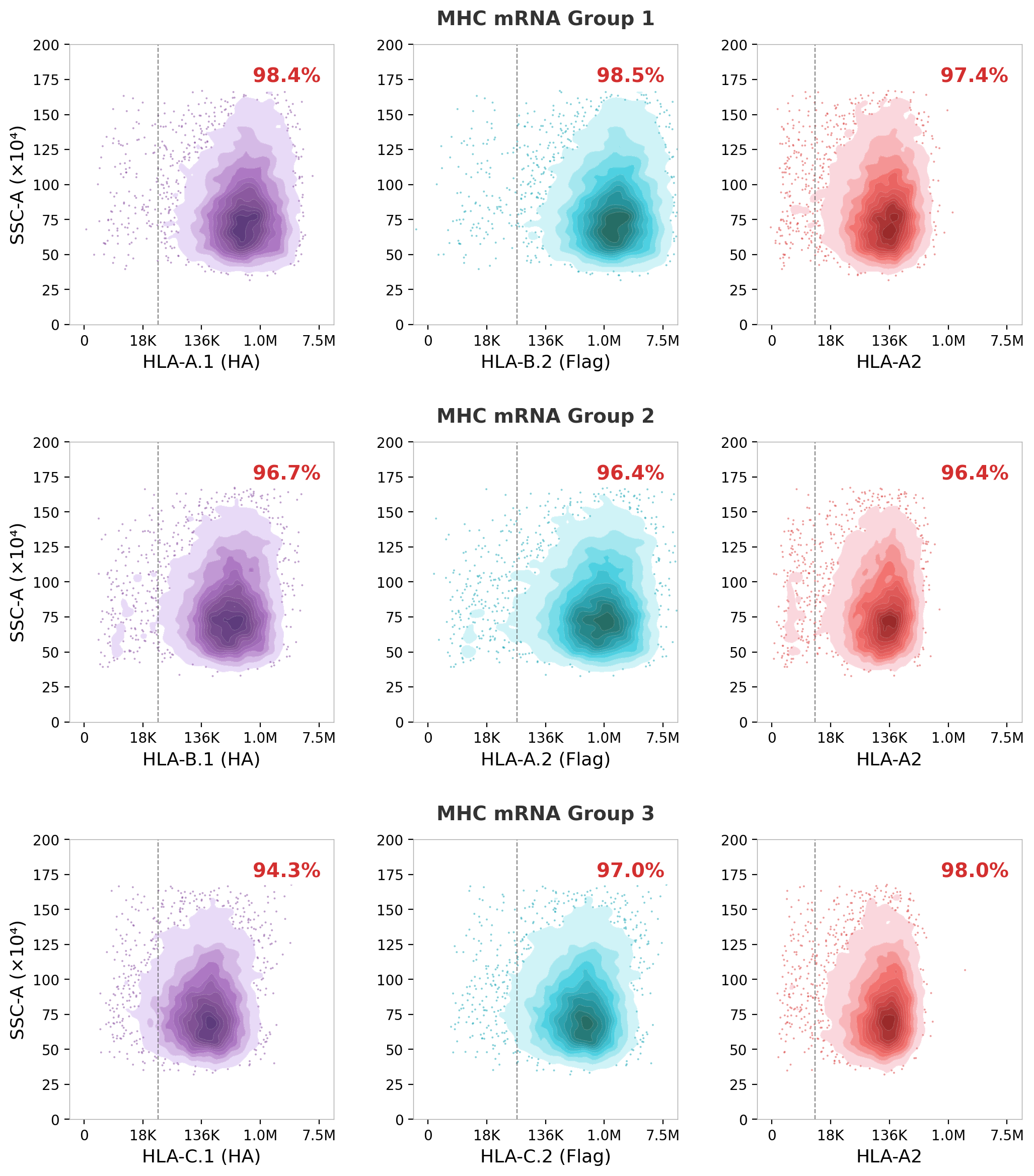
As shown in Figure 3, both anti-HA and anti-Flag stain positive in all 3 preps, although the expression level of the HLA-C alleles still appear somewhat lower than HLA-A and HLA-B. The spike-in HLA-A*02:01 can also be readily detected.
Positive-control pMHC Presentation
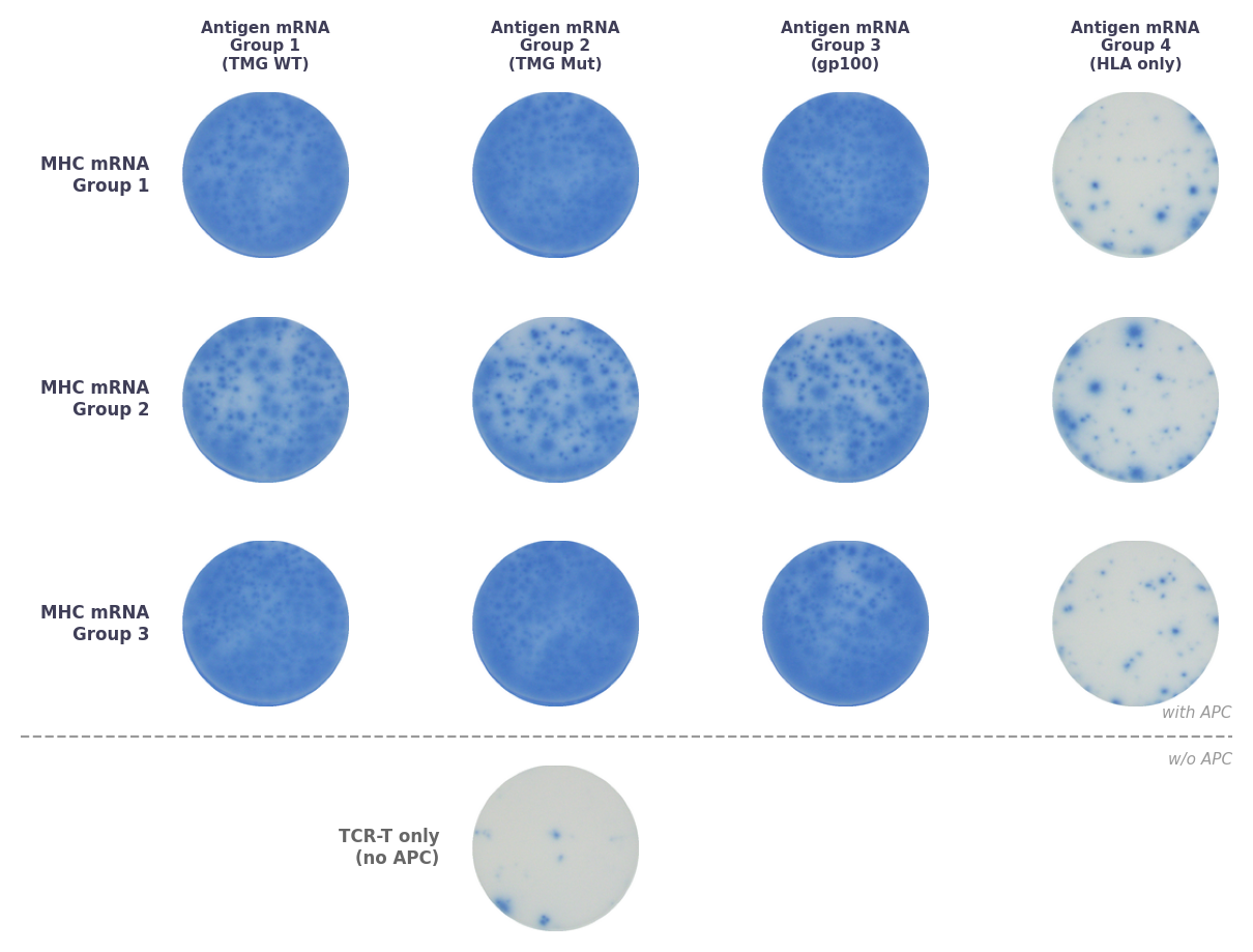
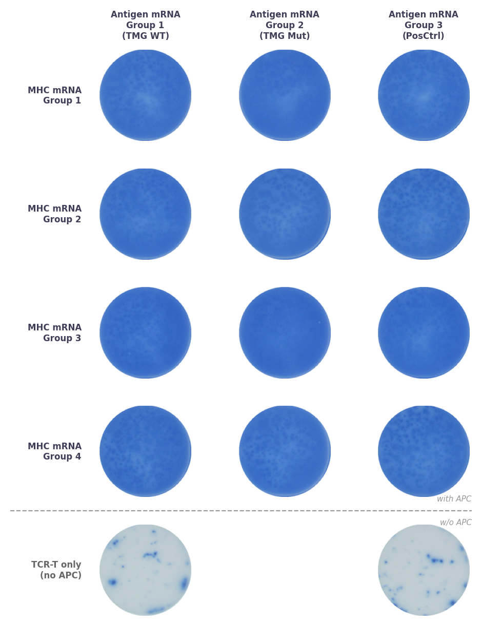
In this example project we used 4 Antigen mRNA Groups:
| Candidate Antigens | Positive-control Antigen | |
|---|---|---|
| Antigen mRNA Group 1 | TMGs of WT sequences | WT-1 epitope |
| Antigen mRNA Group 2 | TMGs of Mut sequences | WT-1 epitope |
| Antigen mRNA Group 3 | gp100 | WT-1 epitope |
| Antigen mRNA Group 4 | None | None |
In the QC step, each Synthetic APC Prep is co-cultured with WT-1-specific TCR-T cell line in an IFN$\gamma$ ELISPOT assay. The grid below shows results. Antigen mRNA Groups 1–3 each include the WT-1 positive-control epitope alongside the candidate antigen, registering strong IFN$\gamma$ signal when co-cultured with WT-1-specific TCR-T. Antigen mRNA Group 4 doesn’t contain any antigen, and as expected, does not activate WT-1-specific TCR-T. WT-1-specific TCR-T cells alone do got generate signal either.
Class II MHC
MHC Surface Expression
The monoallelic nature of ${\beta}2m$ makes Class I MHC relative easy to understand and handle. Class II MHC is considerably more complicated, since each Class II MHC is formed by an HLA Class II $\alpha$ chain and an HLA Class II $\beta$ chain, each of which are polymorphic. HLA Class II $\alpha$ chain is encoded by 3 loci: HLA-DRA, HLA-DQA1, HLA-DPA1. Similarly, HLA Class II $\beta$ chain is encoded by 3 loci: HLA-DRB1, HLA-DQB1, HLA-DPB1. As before, we use the suffix ‘.1’ and ‘.2’ to distinguish two alleles of the same gene. For any isotype (DR, DQ or DP), either $\alpha$ chain may form a dimer with either $\beta$ chain. For example, any of the 4 heterodimers may be present on a patient’s cell surface:
- HLA-DQA1.1 + HLA-DQB1.1
- HLA-DQA1.1 + HLA-DQB1.2
- HLA-DQA1.2 + HLA-DQB1.1
- HLA-DQA1.2 + HLA-DQB1.2
We took a similar approach as Class I, and make sure there is only 1 HLA-DR heterodimer, 1 HLA-DQ heterodimer, and 1 HLA-DP heterodimer in a Synthetic APC Prep, and if the anti-HLA-DR, anti-HLA-DQ, and anti-HLA-DP antibodies all stain positive, we know all three heterodimers are expressed in this prep.
The table below describes a typical grouping strategy.
| DPA1 | DPB1 | DQA1 | DQB1 | DRA | DRB | |
|---|---|---|---|---|---|---|
| MHC mRNA Group 1 | DPA1.1 | DPA1.1 | DQA1.1 | DQA1.1 | DRA | DRB1.1 |
| MHC mRNA Group 2 | DPA1.1 | DPA1.2 | DQA1.1 | DQA1.2 | DRA | DRB1.2 |
| MHC mRNA Group 3 | DPA1.2 | DPA1.1 | DQA1.2 | DQA1.1 | DRA | DRBn.1* |
| MHC mRNA Group 4 | DPA1.2 | DPA1.2 | DQA1.2 | DQA1.2 | DRA | DRBn.2* |
* Since some individuals have additional DRB genes in addition to DRB1, they can be added in Group 3 and Group 4, and named DRBn.1 and DRBn.2 here.
One caveat we have to live with is that, as is the case for Class I, the anti-HLA-DP antibody does not stain all allelic combinations between DPA1 and DPB1. Anti-HLA-DQ and anti-HLA-DR antibodies have the same issue. However, introducing epitope tags such as HA and Flag does not work as universally as in Class I. So in our standard workflow, the Class II HLA genes remain untagged. If, for example, the anti-HLA-DP antibody doesn’t stain the synthetic APC electroporated with a mRNA group that contains a particular DPA1 and DPB1 combination, we will conduct a separate electroporation experiment where we only send in this combination to the K562 cell, and use anti-Class-II-MHC antibody to assess whether this combination express at all, although we still cannot distinguish these two possibilities for the Synthetic mRNA Prep containing multiple isotypes: (1) this combination is not expressed in the fully loaded K562 cell, and (2) this combination is expressed in the fully loaded K562 cell, but the antibody doesn’t stain it.
Similar to the WT-1-based positive control for the Class I MHC and antigens, for Class II, we use a DRA*01:01/DRB1*07:01-restricted neoantigen as a positive control. We include this antigen in each Antigen mRNA Group as well. To distinguish this positive-control HLA-DR and patient-derived HLA-DR, this positive-control DRA*01:01 is tagged with HA, and this positive-control DRB1*07:01 is tagged with Flag. (Although these tags do not work universally, they do work for this heterodimer.)
Let’s use a real case to describe the grouping strategy and troubleshooting process. Here’s the Class II HLA type of a patient:
| Assigned Name | HLA Allele |
|---|---|
| DPA1.1 | DPA1*01:03 |
| DPA1.2 | DPA1*02:01 |
| DPB1.1 | DPB1*04:02 |
| DPB1.2 | DPB1*11:01 |
| DQA1.1 | DQA1*02:01 |
| DQA1.2 | DQA1*05:05 |
| DQB1.1 | DQB1*02:02 |
| DQB1.2 | DQB1*03:01 |
| DRA | DRA*01:01 |
| DRB1.1 | DRB1*07:01 |
| DRB1.2 | DRB1*11:04 |
Here’s the composition of each MHC mRNA Group:
| HLA-DP | HLA-DQ | HLA-DR | Positive Control MHC | |
|---|---|---|---|---|
| MHC mRNA Group 1 | DPA1.1 + DPB1.1 | DQA1.1 + DQB1.1 | DRA + DRB1.1 | None |
| MHC mRNA Group 2 | DPA1.1 + DPB1.2 | DQA1.1 + DQB1.2 | DRA + DRB1.2 | DRA*01:01 (HA) + DRB1*07:01 (Flag) |
| MHC mRNA Group 3 | DPA1.2 + DPB1.1 | DQA1.2 + DQB1.1 | DRA + DRB1.1 | None |
| MHC mRNA Group 4 | DPA1.2 + DPB1.2 | DQA1.2 + DQB1.2 | DRA + DRB1.2 | DRA*01:01 (HA) + DRB1*07:01 (Flag) |
Table 2. Class II MHC mRNA grouping strategy
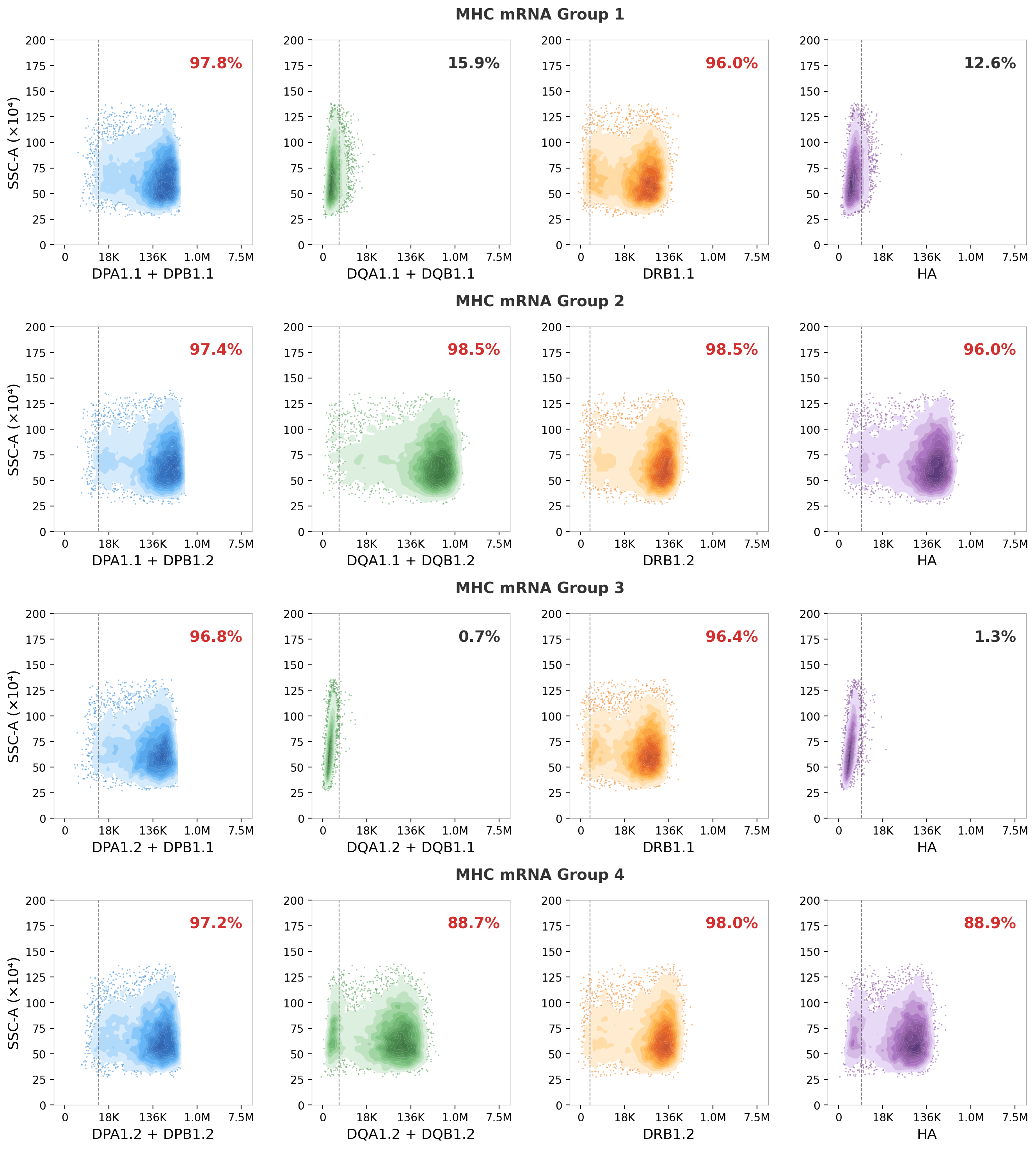
From Figure 5 it can be seen that most staining results are as expected. However, the anti-HLA-DQ antibody fails to detect the DQ heterodimer in Groups 1 and 3, illustrating the antibody coverage limitation discussed above.
The two heterodimers that failed to be stained are DQA1*02:01/DQB1*02:02 and DQA1*05:05/DQB1*02:02. To study whether the negative staining is due to lack of heterodimers on the cell surface or inability of the anti-HLA-DQ-antibody does not stain these heterodimers, we electroporated each combination into K562 cells, and stained with anti-pan-Class-II MHC antibody.
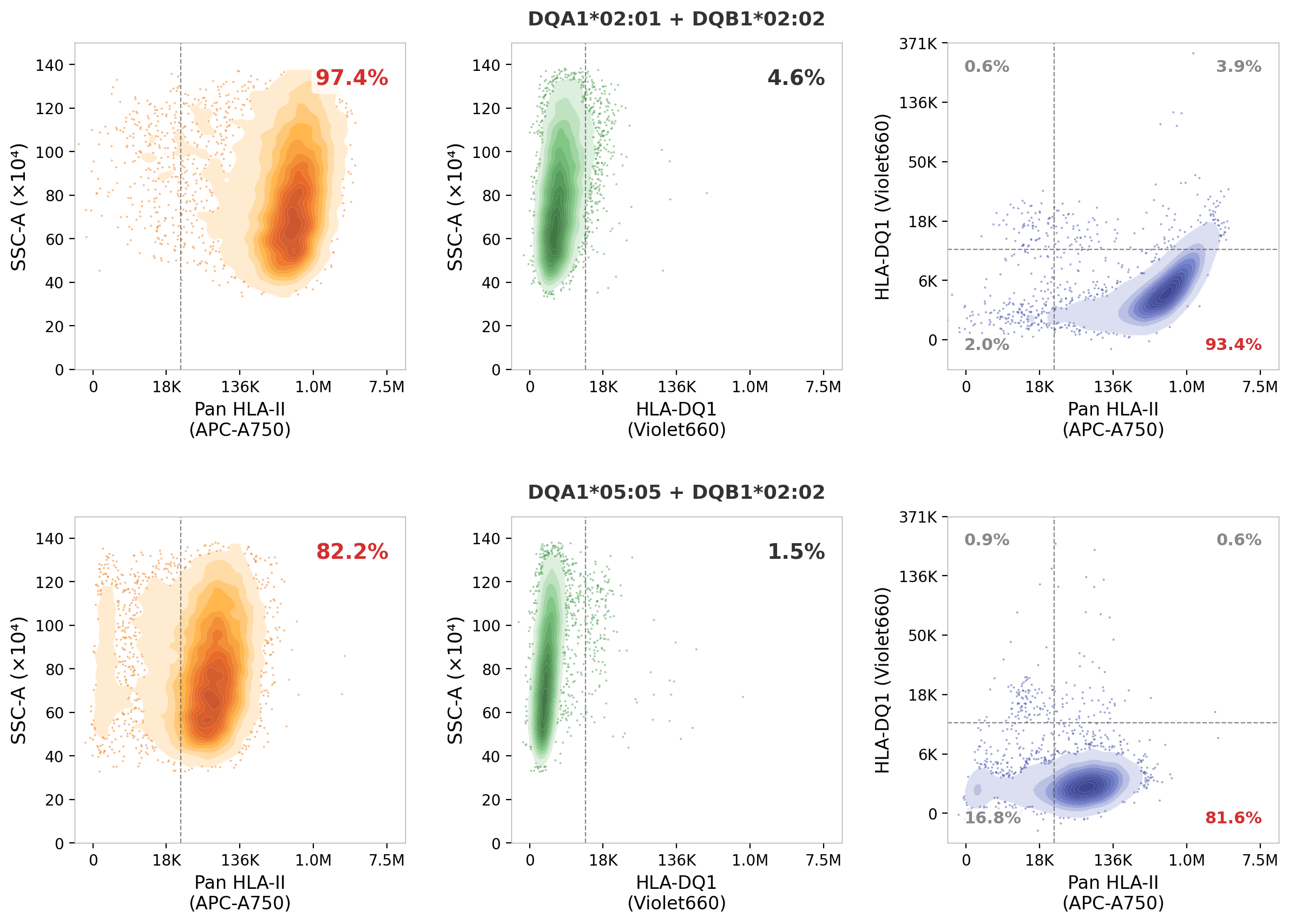
As can be seen on Figure 6, when a single heterodimer is introduced to K562 cells, the anti-pan-Class-II MHC antibody readily stains the cells, indicating that these heterodimers do express, but the anti-HLA-DQ antibody failed to stain them.
Positive-control pMHC Presentation
In this project, we used 2 Antigen mRNA Groups:
| Candidate Antigens | Positive-control Antigen | |
|---|---|---|
| Antigen mRNA Group 1 | TMGs of WT sequences | Mutant CTSB epitope |
| Antigen mRNA Group 2 | TMGs of Mut sequences | Mutant CTSB epitope |
Considering there are 4 MHC mRNA Groups and 2 Antigen mRNA Groups, 8 Synthetic APC Preps was prepared. In QC experiments, each prep was co-cultured with the positive-control TCR-T (NCI4136M, which recognizes mutant CTSB epitope included in both Antigen mRNA Groups).
Conclusion
We have developed a systematic methodology to ensure that we can confirm the expression (on the protein level) of each Class I and Class II MHC molecules on the surface of the synthetic APC. We also use positive-control pMHCs and their corresponding TCR-T cells to ensure that not only the MHC molecules can be detected by antibodies, but the antigen can be properly processed, and the pMHC complex can be recognized by TCR. In the course of providing the TCR Decoder™ service, we have discovered novel phenomena regarding MHC expression. This fully synthetic system not only make the logistics much easier (no need to produce and ship B-LCLs and MDDCs), but also provides great details about MHC biology.
“What I Can’t Create, I Don’t Understand” – Richard Feynman
When we use B-LCL and MDDC, we take it for granted that all six Class I MHCs and all of the 12-ish Class II MHC alleles are expressed and all of the 12-ish Class II MHC heterodimers are formed on the cell surface. Are they, really? Immunologists rarely examine these cell lines for expression or loss of MHC, especially on the protein level. But that’s ok. These tools have long track records of being useful.
However, when we take things into our own hands and express the recombinant MHCs in cell lines such as K562, it’s up to us to prove all expressible MHCs are indeed expressed. This turns out to be more challenging than one may think. First, not all MHC allele has its own antibody with which you can stain the Synthetic APC Preps to confirm expression. The best tool we have on the protein level are isotype-specific antibodies such as HLA-A-, HLA-B- and HLA-C-specific antibodies. But if we express two HLA alleles (e.g., HLA-A*02:01 and HLA-A*11:01), and stain with this antibody, how do we know which one is expressed? We will spend the bulk of this post addressing this problem. But let me offer an interesting observation first. If we express an HLA-A, and HLA-B and HLA-C allele in K562 cells, and stain the cell with HLA-A-, HLA-B-, and HLA-C-specific antibodies, we found that the HLA-C has very low expression level, almost undetectable. But when we express HLA-C alone in K562, it can express fairly well.
Is this inter-HLA-competition and inhibition biologically meaningful? I think it’s meaningful in at least some context. But importantly, building the synthetic platform obliges us to perform such characterization, leading to a much higher level of clarity when studying systems as complex as immunology.
– Xi Chen